HESA Committee Report
If you have any questions or comments regarding the accessibility of this publication, please contact us at accessible@parl.gc.ca.
Appendix A: Photographs of Breast Implant Complications
WARNING: This appendix contains images with partial nudity and graphic medical content.
Patient advocates have recommended that Health Canada include photographs showing the complications of breast implants on its website to increase awareness of potential adverse effects and improve informed consent. Figures A1 through A10 provide a selection of such photographs.
Figure A1—Swollen Left Breast in a Patient with Breast Implant-Associated Anaplastic Large Cell Lymphoma (BIA-ALCL)

Note: The patient’s left breast is swollen. One of the main symptoms of BIA-ALCL is persistent swelling around the breast implant.[1]
Source: Photo courtesy of the Breast Implant Failure & Illness Society – Canada, submitted to the committee by Nancy Pratt.
Figure A2—Syringes Filled with Seroma from a Patient Diagnosed with BIA‑ALCL
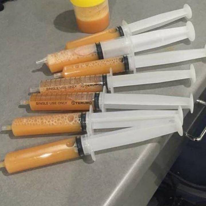
Note: Patients with BIA-ALCL often have an accumulation of fluid (seroma) around the breast implant.[2]
Source: Photo courtesy of the Breast Implant Failure & Illness Society – Canada, submitted to the committee by Nancy Pratt.
Figure A3—Mass Found in Capsule of Patient with Stage 4 BIA-ALCL
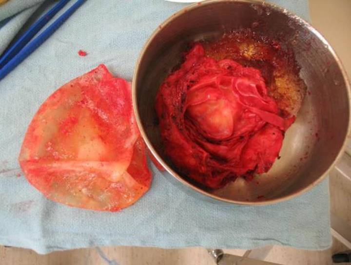
Note: Lumps around the breast or armpit can be a sign of BIA-ALCL.[3]
Source: Photo courtesy of the Breast Implant Failure & Illness Society – Canada, submitted to the committee by Nancy Pratt.
Figure A4—Ruptured Cohesive Silicone Gel (“Gummy Bear”) Breast Implants
Note: Rupture is a possible complication of both saline and silicone implants.
Source: Photo courtesy of Dr. Aditya Sood, submitted to the committee by Nancy Pratt.
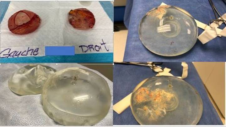
Figure A5—Rupture of Poly Implant Prothèse (PIP) Breast Implant
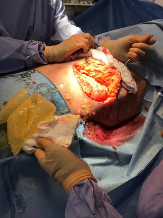
Note: Silicone implants manufactured by the French company Poly Implant Prothèse (PIP) were removed from the market after health authorities in France discovered they contained industrial‑grade rather than medical-grade silicone and were more prone to rupture.[4] Although PIP implants were never distributed in Canada, patients in Canada may have received them while abroad.
Source: Photo courtesy of Dr. Stephen Nicolaidis, submitted to the committee by Nancy Pratt.
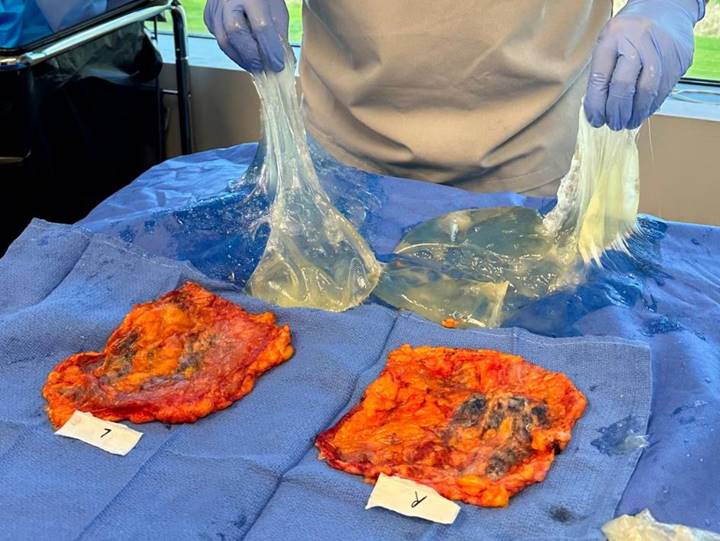
Figure A6—Silicone Granulomas
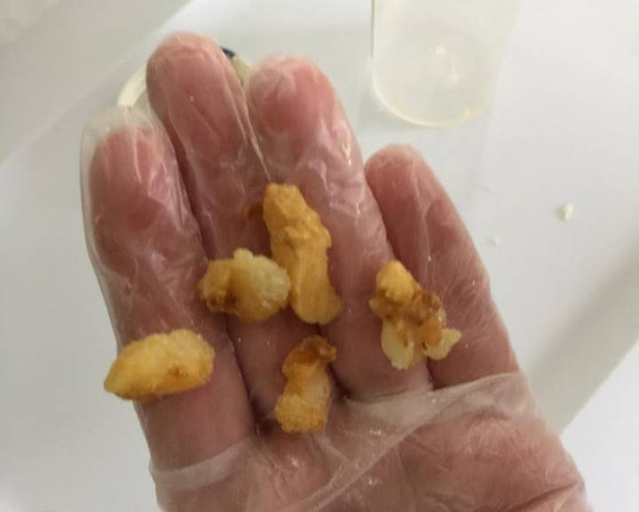
Note: Silicone granuloma formation may occur following extracapsular rupture of a breast implant (i.e., when silicone escapes beyond the capsule around the implant). There have been reports of silicone granulomas found in numerous sites throughout patients’ bodies.[5]
Source: Photo courtesy of the Breast Implant Failure & Illness Society – Canada, submitted to the committee by Nancy Pratt.
Figure A7—Debris in a 23-Year-Old Saline Breast Implant
Source: Photo courtesy of Dr. Aditya Sood, submitted to the committee by Nancy Pratt.
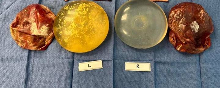
Figure A8—Smooth Saline Implants Recalled Because of Valve Issue
Note: The top left image shows implants in scar capsules. The bottom left image shows a saline leak. The photos on the right show discolouration with possible contamination of the implant.
Source: Photo courtesy of the Breast Implant Failure & Illness Society – Canada, submitted to the committee by Nancy Pratt.
Figure A9—Grade IV Capsular Contracture
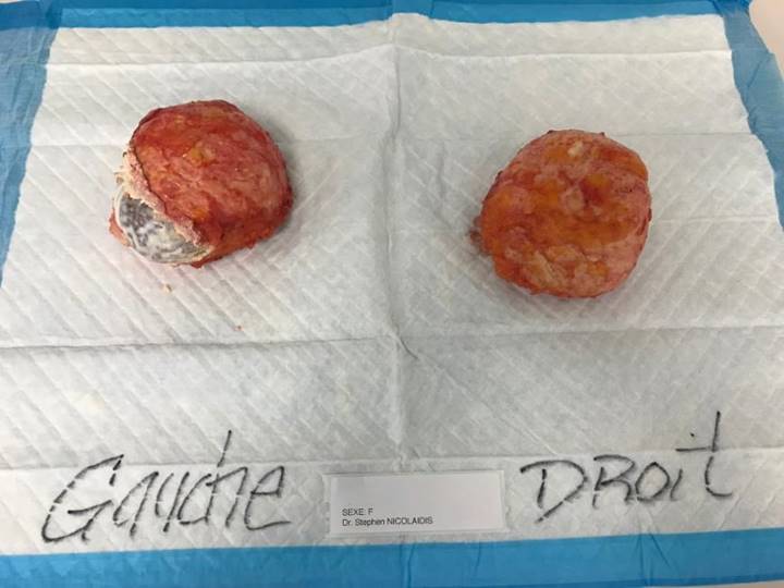
Note: Capsular contracture refers to the tightening or hardening of scar tissue around the implant. The severity of capsular contracture is graded on a scale from I to IV, with I representing the least severe (asymptomatic) and IV representing the most severe (hard, misshapen and/or painful breasts).[6]
Source: Photo courtesy of Dr. Stephen Nicolaidis, submitted to the committee by Nancy Pratt.
Figure A10—Heavily Calcified 30-Year-Old Breast Implants
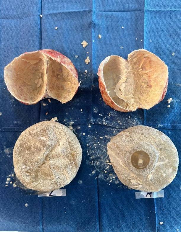
Note: Calcium deposits can form on the breast implant capsule. Calcification can interfere with breast cancer screening.[7]
Source: Photo courtesy of Dr. Aditya Sood, submitted to the committee by Nancy Pratt.
[1] United States Food and Drug Administration, Questions and Answers about Breast Implant-Associated Anaplastic Large Cell Lymphoma (BIA-ALCL).
[2] United States Food and Drug Administration, Questions and Answers about Breast Implant-Associated Anaplastic Large Cell Lymphoma (BIA-ALCL).
[3] United States Food and Drug Administration, Questions and Answers about Breast Implant-Associated Anaplastic Large Cell Lymphoma (BIA-ALCL).
[4] France 24, French PIP breast implants: an ongoing global health scandal, 29 September 2018.
[5] Hanad Ahmed et al., “Chest Wall Silicone Granuloma Following Ruptured Silicone Breast Implant Causes Giant Chest Wall Abscess and Osteomyelitis: The First Report,” European Journal of Breast Health, Vol. 17, No. 4, October 2021.
[6] Kevin Tehrani, “What is capsular contracture and how can it be treated?,” American Society of Plastic Surgeons, 12 June 2018.
[7] United States Food and Drug Administration, Risks and Complications of Breast Implants.